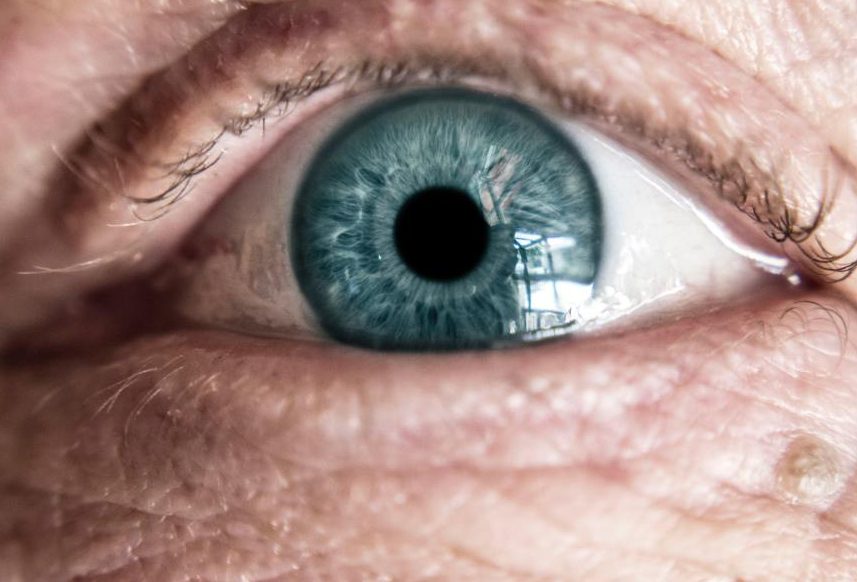
Vision damage can occur after the age 40.
If caught early, some of these conditions can be treated without irreversible damage to the vision. Others can be improved with early treatment and can last a lifetime.
For this reason, a detailed ophthalmological check-up is required after the age of 40, once a (normal) physiological phenomenon occurs – the decrease in reading vision (presbyopia/presbyopia, which requires an optical correction).
These conditions include glaucoma and age-related macular damage (AMD).
Glaucoma can be a group eye disease that is often linked to an increase in intraocular Pressure. This can cause progressive damage and loss of nerve fibers. Another theory is that there is not enough blood supply to optic nerve. Blindness can result from untreated or poorly diagnosed glaucoma.
Primitive open angle glaucoma (GPUD), which is most common in older people, develops slowly and usually without symptoms. It is often confused with the need to wear glasses because it occurs with the onset or presbyopia (decreased ability to read). It is important to have an ophthalmological exam after 40. This disease is often not recognized by many people until they experience significant vision loss. It can affect peripheral vision initially, but can lead to central vision loss. Glaucoma, if left untreated can cause significant vision loss in both of the eyes.
Risk factors include:
- Age – People over 40 are at greater risk of developing the disease
- Race – African Americans are more vulnerable than Caucasians.
- Family history of glaucoma
- medical conditions like diabetes, hypertension, heart diseases, etc
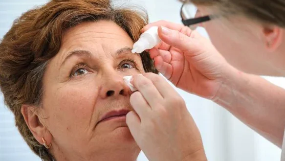
- A thorough eye exam is required to diagnose glaucoma.
The following are examples of testing:
- The patient’s medical history
- Measurement of visual acuity
- Intraocular pressure measurement
- Measure the thickness of your cornea
- Visual field testing
- fundus examination
- Examination of the chamber angle. OCT (optical coherent tomography) of optic nerve. A few optic nerves can have a suspicious appearance similar to glaucoma. However, patients do not have any other signs or risk factors. Routine eye exams should be performed on these patients to monitor their progress.
Glaucoma treatment is designed to lower intraocular pressure and increase arterial circulation to the optic nerve. Prescription eye drops must be used regularly as the first treatment. Sometimes, surgery, laser treatment or systemic drugs may be required. When other treatment options are ineffective, surgery may be necessary. This can be either the traditional method (trabeculectomy and peripheral iridectomy), or it may involve the placement of a small device that drains the aqueous humor to lower intraocular pressure (ex.Press).
Acute angle-closure is a less common form of glaucoma. It occurs when intraocular pressure rises rapidly. This condition can cause severe eye pain, nausea and blurred vision. This is an emergency situation that requires immediate medical attention. It can cause severe vision loss and can be controlled by prompt treatment.
Age-related macular disease (AMD), which affects people over 50, is characterized with decreased central vision and/or metamorphopsia, a distortion of the perception of images. The central part of the retina responsible for seeing colors and details is called the macula.
DMLV-Classification:
- Dry (atrophic), 80% of cases The patient may not notice the loss of central sight until it is too late.
- 20% of cases are wet (exudative). It is more serious and progresses quicker.
Symptoms of DMLV
- Initial stage: gradually decreasing vision (with reading difficulties); vision “in fog”; poor color perception; low adaption to changes in brightness;
- decreased contrast capacity; straight lines appear distorted
- Advanced stage: The central portion of the visual fields has been lost; the patient can see a dark spot in their visual field.
DMLV risk factors:
- Predetermined factors: Age (over 50), gender (female); DMLV in the family; low cell density in the macula
- Factors that can be affected include smoking, poor diet rich in vitamins, Omega 3 and mineral intake, obesity, arterial hypertension, increased blood lipids; coronary disease; UV exposure.
Diagnosis for DMLV:
- Only after an ophthalmological consultatio
- The Amsler grid can help the patient detect the early stages of disease.
DMLV-Classification:
- Dry (atrophic), 80% of cases The patient may not notice the loss of central sight until it is too late.
- 20% of cases are wet (exudative). It is more serious and progresses quicker.
Symptoms of DMLV
- Initial stage: gradually decreasing vision (with reading difficulties); vision “in fog”; poor color perception; low adaption to changes in brightness; decreased contrast capacity; straight lines appear distorted
- Advanced stage: The central portion of the visual fields has been lost; the patient can see a dark spot in their visual field.
DMLV risk factors:
- Predetermined factors: Age (over 50), gender (female); DMLV in the family; low cell density in the macula
- Factors that can be affected include smoking, poor diet rich in vitamins, Omega 3 and mineral intake, obesity, arterial hypertension, increased blood lipids; coronary disease; UV exposure.
Diagnosis for DMLV:
- Only after an ophthalmological consultation
- The Amsler grid can help the patient detect the early stages of disease.
DMLV treatment consists in a healthy diet that focuses on raw fruits and vegetables, as well as drug treatment (rich with lutein, zeaxanthin and Omega 3 fatty acid, etc.). As indicated by the ophthalmologist. Intravitreal injections of anti-VEGF drugs, etc.
Cataract refers to the alteration in transparency of the crystal lens. It can be congenital, acquired or both.